Uretero-renoscopes
High-contrast images with high stability
Ureterorenoscopy allows the visualization of the bladder, ureter, and renal pelvis. In addition to diagnostic purposes, it is also used for treatment. The basis of this application are the uretero-renoscopes. These endoscopes with working and irrigation channels have a high-quality, contrast-enhancing image bundle design. This provides users with both a good image and a high level of stability.
High-quality, contrast-enhancing image bundle as a quality feature
High-quality image bundles ensure optimal image transmission. Our uretero-renoscopes also have low service requirements and a long service life due to the image bundle design. They achieve a homogeneous image with a low grid structure.
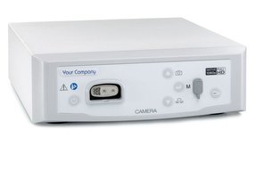
The Grid Removal video algorithm of the FlexiVision camera series can also optimize the image by reducing the visibility of the grid structure as much as possible.

Durability and high comfort, flexibility in use as a quality feature
A bending radius of 15° in all directions offers good handling. This enables users to optimally follow the anatomical conditions without risking a defect.
A lithotripter can be inserted with the URS through the straight working channel. This design also facilitates handling and exchange of instruments during surgery.
Different instruments such as laser fibers, lithotripters or stone baskets can be used.

Atraumatic tip element as a quality feature
The gently stepped increase in the outer diameter allows for atraumatic insertion and gentle stretching of the tissue.

Removable instrument bridge as a quality feature
The removable instrument bridge simplifies and improves the cleaning and reprocessing of the uretero-renoscopes.
Uretero-renoscopes and instrument bridges form a closed system that cannot be combined with bridges from other manufacturers. This ensures that unsuitable or inadequate instrument bridges cannot be connected.


