Visual testing during stents production
Optical inspection during series production of vessel supports
Stents inserted into coronary vessels have a vital function for humans. They are subject to strict quality controls during production. Visual testing can be carried out using the inspection system FlexiVision 100 and a special SCHÖLLY device.

Non-destructive testing of fine structures
Stents are used in the medical field to reopen narrowed coronary vessels and allow undisturbed blood flow. To ensure that the stents function properly, visual testing is carried out during production. The structure of the stent is created by laser cutting. For this purpose, a filigree metal tube is processed with the laser. This gives the stent a specific structure that later resembles a grid when open.

Detecting errors from laser cutting by visual testing
During the visual inspection in production, the result of the laser cutting is checked. Due to light reflections of the metal, conventional visual testing by means of a borescope and illumination from the inside is not effective. The stent is therefore illuminated from the outside, which causes a certain shadow image of the metal. This image is checked segment by segment with an unlit borescope. Fractures or errors from laser cutting are thus detected.

Tools for optimized visual testing
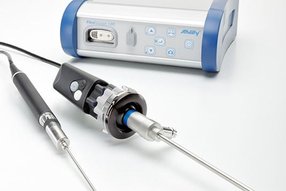
For this inspection procedure, SCHÖLLY developed a device in which the visual inspection takes place inside a heat sink in order not to damage the sensitive material and to illuminate the stent uniformly from the outside. The testing device with integrated borescope and ring illumination with diffuser is connected to a light source and to the inspection system FlexiVision 100. The test images are transmitted to a monitor. The borescope with a diameter of 0.9 mm and a viewing direction of 7.5° was specially developed for this application. The outer diameter of the stents is between 1.6 and 2.3 mm.

Visual inspection of opened stents
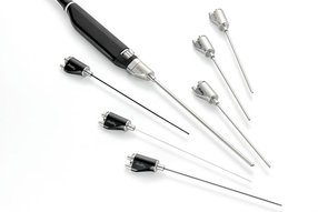
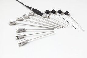
In addition to closed stents, opened stents are also tested by random sampling. Here the quality of the structure is checked optically. With the inspection system FlexiVision 100 and the associated camera handpiece, various probes from a diameter of 0.7 mm can be used for this inspection. With the high resolution of the camera (1920 x 1080p) the fine structure of the stent can be easily visualized and evaluated.

Visual inspection with FlexiVision 100
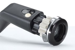
The device enables the employee to generate optimal images for quality inspection. By feeding the light from the outside and the special viewing direction, he can very well recognize the structures of the closed stent and immediately detect possible fractures or defects. This allows the optical visual inspection to be carried out faster and more reliably than with conventional methods. With the connection of the device and the FlexiVision 100, the inspector can also use many other functions. If required, the inspector can, for example, store the inspection images on a USB stick at the touch of a button. During the inspection process it is possible to position a reference image or an error image on the screen (split screen function). Thus a direct comparison can be made during the visual inspection.