 Open video in modal
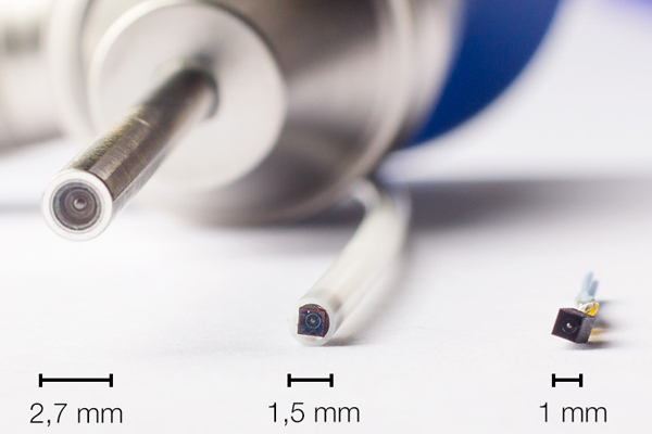
Very small image sensors
Using very small image sensors in CIT systems means that there is more space for other functions.
For applications such as uretero-renoscopy, endoscopes with integrated working channels are used. If the space required for image transmission is reduced by a smaller chip at the tip, other required functions such as the working channel for instruments or the flow for suction can be increased.

Miniaturized visualization systems enable previously invisible areas to be seen
New areas of application can be opened up by installing the image sensor so that it can be inserted through a vessel coated by a flexible hose. Applications such as in cardiology are just as conceivable here as miniaturized visualization systems for endoscopic spinal surgery or sialoendoscopy.
How is the image quality of thin scopes improved?
The combination of CIT endoscopes with a flexibly configurable camera platform enables image quality to be improved. By adapting the camera precisely to the connected endoscope and optimally adjusting the video algorithms to it, the image quality can be improved even with the smallest endoscope diameters.
Patents
SCHÖLLY has patented various ideas in the field of 2D/3D CIT endoscopes. More information is available here.